Allografts
Bone allografts are available for use in orthopaedic and dental surgeries. Although autograft bone is considered a gold standard, in many clinical cases the use of autograft is not always necessary and allograft can serve the same purpose. Allografts are available off the shelf, save time and cost to harvest, avoid donor site morbidity, available in unlimited quantity and can also be mixed with autograft in certain challenging cases such as revision surgeries. Allografts come in 3-4 year shelf life and stored at room temperature, ready for use. There are two types of bone allografts; osteoconductive Cancellous Chips for filling bone defects and osteoinductive (new bone forming) Demineralised Bone Matrix (DBM) for fusing bone and joints. Chips may be mixed with DBM as a graft extender in larger bone defects. The following table shows the types of bone grafts and their properties.
Slide right to see full table as required
| Bone graft | Osteoconduction | Osteoinduction | Osteogenesis |
| Autograft | +++ | +++ | +++ |
| Autograft Cancellous Chips | +++ | - | - |
| Allograft DBM | + | +++ | - |
| Synthetic bone | ++ | - | - |
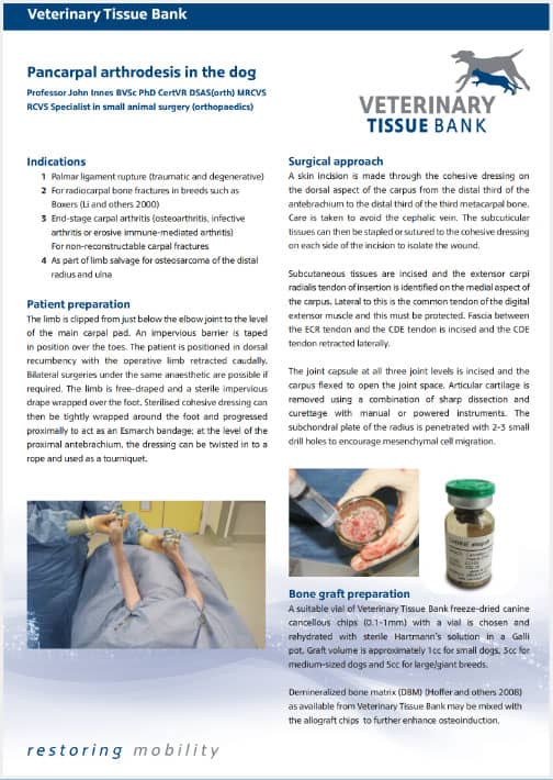
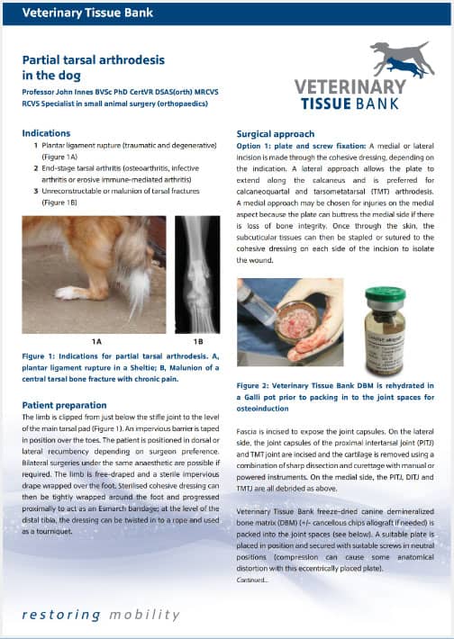
Allograft Chips and DBM are available in 1cc, 3cc and 5cc. For most cases 1cc is sufficient such as in arthrodesis to fill in metacarpal or tarsal joints in dogs. The following table shows the indications for various allograft bone in dogs and cats.
| Type | Indications |
| DBM | Fusion of joints in athrodesis, spinal fusion, non-union or delayed union, revision surgeries, periodontal use |
| Cancellous Chips | Filling in bone cysts, acute fractures, graft extender to DBM |
| Cortical segment | Spinal fusion, tumour resection |
| Cortical strut | Augmentation of long bone |
| DBM putty | IOHC (with IOHC screw), fusion of joints |
Demineralised Bone Matrix (DBM)
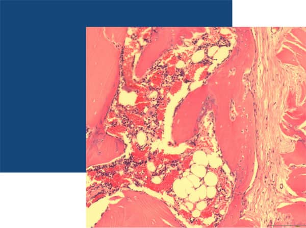
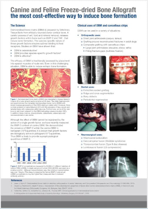
Demineralised Bone Matrix (DBM) DBM is prepared from cortical bone which contains osteoinductive growth factors (BMPs) including BMP2 and BMP7 which are relevant to bone healing. DBM remains the only type of bone graft which contains growth factors and therefore superior to synthetic bone grafts which act as osteoconductive filler. The presence of BMP can be demonstrated by implanting DBM in vivo and examined under microscope for new bone formation. The figure below shows the H&E stained DBM after 28 days of implantation in thigh muscle of an athymic mouse, showing new bone forming elements such as osteocytes, adipocytes, bone marrow, new osteons, etc.